Love & Orthodontics
  • Love & Orthodontics
  • Online Courses
  • Enroll
  • Love & Ortho Blog
  • Love & Orthodontics
  • Online Courses
  • Enroll
  • Love & Ortho Blog

The Hidden Secrets the Panoramic Holds

5/24/2018

1 Comment

 
Picture
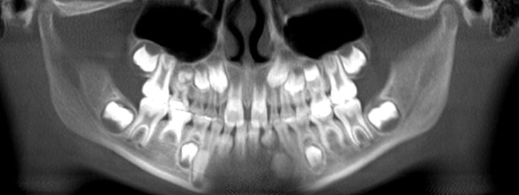
Missing upper 2's, missing lower 5's, ectopic canines. © 2018 Dr Chris Baker
The Panoramic radiograph.  You may be surprised to discover the amazing information you can find about a child patient in that x-ray.  Can you name five really important findings in a panoramic on an early mixed dentition patient?

Can you name six?  Seven?  Let’s go for double that figure! (And we won’t include decay, as it’s not the best diagnostic tool of decay.  Well, on second thought, maybe we will include some  possible decay-related findings at the end of our discussion.)

The panoramic should be taken when any permanent teeth are erupting.  Often those first eruptions are the lower incisors, or the six-year molars, but no matter what teeth, take the pano then.  (You may find opportunity to change the child’s life because of the secrets that x-ray holds.)

Start by counting/identifying all teeth (Sounds pretty tame and familiar so far!) :
  1. Missing teeth.  Count ‘em.  Every single one.  Be sure.  Most often upper lateral incisors, and bicuspids, but maybe any incisor, any tooth actually.  While severe “anodontia” is typically related to Ectodermal Dysplasia, and other developmental disorders, any permanent tooth can be missing even without identifiable developmental disorders "with names.”
  2. Extra teeth.  Supernumerary teeth can be anywhere.  One of my patients had one that looked like a canine, but about the size of a primary canine, in the floor of the nose, over the bicuspid root area. 
  3. Malformed  and unusual teeth.   While ectopic and unusual trajectories of eruptive paths can cause dilacerations, there may be malformed crowns as well.  Note any taurodontic primary molars that may hinder exfoliation, dilacerated roots that may hinder eruption, or if the dilacerated root is next to a missing tooth space, it could prevent implant later.  Notice now.  Notice incisors with talon lingual cusps/dens evaginates, and shovel-shaped incisors with scooped out lingual surfaces which may be more at risk of decay at the cuspal interfaces, and may create occlusal issues in contact with the lower incisors.  
  4. Variations in sizes of teeth. Notice particularly large lateral incisors as well as peg or small laterals which may also interfere with occlusal harmony.
  5. Fused or possibly fused teeth; or geminated teeth.
  6. Ectopic teeth positions, which can portend huge difficulties in getting the teeth into the arch later, or even contribute to transpositions of teeth, and certainly to extended orthodontic treatment.  If normal eruption does not occur, ectopic teeth may need to be orthodontically moved over larger than normal distances, and past other tooth roots.  This predisposes both the ectopic teeth as well as the teeth in the path of the movement to potential damage, including root resorption, loss of vitality, alveolar bone loss and gingival and per problems, and some ankylose,
  7. Stage of exfoliation and eruption
  8. Poorly resorbing roots on primary teeth - is it due to ectopic position of succedaneous permanent tooth?  Is the permanent successor missing?
  9. Submerged primary molars - actually probably due to an ankylosis or other failure of the tooth to undergo normal eruptive growth with the adjacent teeth.
  10. Crowding.  Eruption issues to come.  Opportunity to clear the path of eruption and prevent more complex problems and need for extended treatments.  
  11. Third molar positions.  While not usually evident in the earliest mixed dentition panoramic x-ray, often are notable within a year or two.  Horizontal and severe ectopic positions may affect the eruption of the twelve year molars, and should be monitored for need for early removal to allow eruption of the twelve year molars.
  12. Twelve year molars that do not have the space to erupt in the mandible.  As human jaws get smaller, and teeth do not, we are seeing more 12 year molars that will never be able to clear the mandibular ramus, like many third molars.  Not frequent, but more and more so.
  13. Class II or III occlusion (at least as screening, to be evaluated further.)
  14. Atypical incisive papilla alveolar bone area.  Again, screening, to be confirmed with diastema, low and deep pull frenum, etc.
There you have some great secrets the pano may hold.  Nice not to be surprised later when it is later, and things have gotten way complex.  Think about what you can do for your patient when you take panos at the time of eruption of permanent teeth. 

Here’s an important opportunity to do outstanding diagnosis for your patient, and be paid for your care.  You’re in dentistry to care for people, and in business to make a living.  
Picture
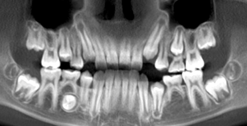
Missing LL5, and severely ectopic LR 5, which had to be removed. © 2018 Dr Chris Baker

Picture
1 Comment
MckimmeCue link
6/6/2022 12:38:29 pm

This is a very informative—edifying article to all. Thanks a lot! Continue to post!
If you are looking for coupon codes and deals just visit coupon plus deals dot com

Reply


Leave a Reply.

    Picture

    Dr Chris Baker

    America's most-trusted teacher of orthodontic continuing education, Dr. Chris Baker has practiced and taught for more than 30 years, and is a current or former faculty member of three U.S. dental schools.  She is a pediatric dentist, author, blogger, dental practice consultant, and mentor.  Dr. Chris is also Past President and Senior Instructor of the American Orthodontic Society.  She is based in Texas, USA, but lectures around the world.

    Categories

    All
    Abu Dhabi To Texas
    Dental Practice Freedom
    Life & Home
    L & O TV
    Orthodontics
    Pediatric Dentistry
    REAL Brief

    Archives

    January 2023
    December 2022
    November 2022
    October 2022
    September 2022
    August 2022
    July 2022
    June 2022
    May 2022
    April 2022
    March 2022
    February 2022
    January 2022
    December 2021
    November 2021
    October 2021
    September 2021
    August 2021
    July 2021
    June 2021
    May 2021
    April 2021
    March 2021
    February 2021
    January 2021
    December 2020
    November 2020
    October 2020
    September 2020
    August 2020
    July 2020
    June 2020
    May 2020
    April 2020
    March 2020
    February 2020
    January 2020
    December 2019
    November 2019
    October 2019
    September 2019
    August 2019
    July 2019
    June 2019
    May 2019
    April 2019
    March 2019
    February 2019
    January 2019
    December 2018
    November 2018
    October 2018
    September 2018
    August 2018
    July 2018
    June 2018
    May 2018
    April 2018
    March 2018
    February 2018
    January 2018
    December 2017
    November 2017
    October 2017
    September 2017
    August 2017
    July 2017
    June 2017
    May 2017
    April 2017
    March 2017
    February 2017
    January 2017
    December 2016
    November 2016
    October 2016

    RSS Feed

    Text and images
    © 2022 Dr Chris Baker